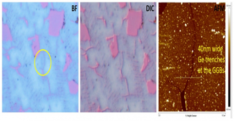
Optical wide-field imaging of sub-diffraction limit nanostructures is of interest in a wide array of applications. In applications where the nanostructures to be visualized are well isolated, a high enough optical contrast is sufficient to detect these. We developed a technique called the Bright-field Nanoscopy which allowed the visualisation of Graphene Grain Boundaries (GGBs), nanoparticles, single isolated Carbon Nanotubes (CNTs). Such remarkable detection was made possible by a device consisting of ultra-thin films of Germanium on optically thick gold substrates. This remarkably low lateral length scale was imaged due to the using of a special thin film structure consisting of a water-soluble thin film layer deposited on a metal substrate, which produces a strong color change as a function of the film thickness. Small local water transport differences in the graphene layer result in thickness variation of the underlying thin film due to its solubility in water and produces color contrast readily observable under a normal bright-field optical microscope with the naked eye. The local color contrast around the nanostructures arise due to difference in water